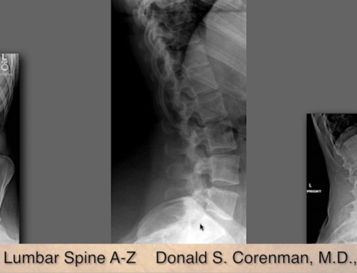
This video: Understanding an MRI of the Normal Lumbar Spine, is designed for the primary care physician or specialist such as a Chiropractor or Physical Therapist to use to learn how to read and understand the MRI of the lumbar spine. The scan shown in this video is normal, meaning, all the structures scanned are normal in appearance and not injured or degenerative. When you can recognize what normal looks like, you can determine what an abnormal finding is. This is the companion video to the next two videos in the series: MRI of lumbar herniated disc and MRI of a lumbar degenerative spondylolysthesis with spinal stenosis. Now-obviously you will not become a musculoskeletal radiologist and be able to identify an abdominal aortic aneurysm by reviewing this video. It is designed to give you a basic understanding of the anatomy of the lumbar spine and then to identify the basic changes that occur with degenerative disc disease, herniated discs and spinal stenosis.
Share This Story, Choose Your Platform!
About the Author: Donald Corenman, MD, DC
Donald Corenman, MD, DC is a highly-regarded spine surgeon, considered an expert in the area of neck and back pain. Trained as both a Medical Doctor and Doctor of Chiropractic, Dr. Corenman earned academic appointments as Clinical Assistant Professor and Assistant Professor of Orthopaedic Surgery at the University of Colorado Health Sciences Center, and his research on spine surgery and rehabilitation has resulted in the publication of multiple peer-reviewed articles and two books.
