An Overview of Isolated Disc Resorption (IDR) of the Lumbar Spine
Isolated disc resorption (IDR) of the lumbar spine is the end spectrum of degenerative disc disease of the lumbar spine. Initially, disc wall tears herald the start of degenerative disc disease. Eventually, the nucleus (the jelly of the donut) loses its ability to hold water and the disc then settles (drops in height). This is similar to a car tire losing air and the walls of the tire bulge out.
The increased pressure on the walls of the disc produces increased stress. The additional forces on this degenerative disc include shear, compressive and rotational forces. More tears can occur over time and the disc can become increasingly incompetent. These abnormal forces can start to wear out all the internal components of the disc including the cartilaginous endplates. Eventually in some discs, this degeneration causes all of these materials (nucleus and endplate) to fully wear away.
Open one of these isolated disc resorption discs and you will find nothing in the disc space but raw bone surfaces. The material within these discs has absorbed away. The annulus remains, normally torn but generally intact.
These changes in the structural dynamics cause symptoms different than the typical back pain most of us know. In the IDR disc, there is no shock absorption capability remaining and the two bony vertebral endplates are set against each other. This creates a real problem with exposure to vibration and impact forces.
Prolonged exposure to vibration from riding in a car or from an airplane ride, impact activities such as jumping, running and tennis will cause pain. Something as simple as stepping off an unexpectedly steep curb will cause a sharp, deep pain in the back. Heavy lifting or prolonged bending will also cause the same type of pain but of a more dull nature.
The isolated disc resorption (IDR) disc will also cause delayed onset pain. This is pain that occurs from 4-12 hours after an overload activity. Most patients note that with loading or impact activities, they “pay for it later”. The quality of this delayed onset pain is an intense dull throbbing ache.
This delayed pain is caused by fractures that occur within the vertebral body in small structural web-like bones called the trabecula. These tiny bones are like a honeycomb inside the vertebra that reinforces the walls. The trabecula are normally protected by the shock absorption capacity of the disc.
When the disc fails, the increased forces on the endplates overcome the structural integrity of these trabecula. When they fracture, raw bone is exposed. This triggers the inflammatory cascade.
The delayed timing of pain is due to the nature of the inflammatory cascade. This chemical cascade is set off when exposed proteins trigger certain immune cells to release their chemical signals. These chemicals in turn cause other cells to migrate into the area and trigger more chemical releases. This cascade eventually sensitizes the nociceptors (pain receptors).
This cascade “revs up” slowly. It takes about 6-12 hours for a cut on your finger to develop red, inflamed edges. This is the same delayed type of reaction that occurs in the spine. There are multiple chemical reactions that have to sequentially complete to fully activate inflammation and induce pain provocation.
Are you suffering from symptoms of Isolated Disc Resorption (IDR) of the lumbar spine?
Would you like to consult with Dr. Corenman about your condition?
You can set up a long distance consultation to discuss your
current X-rays and/or MRIs for a clinical case review.
(Please keep reading below for more information on this condition.)
MRI and CT Findings of IDR
The MRI findings of isolated disc resorption (IDR) are classic. First, the disc will be very narrowed and have lost most of its original height. The disc space will appear black due to the loss of water from the nucleus and then loss of the nucleus itself.
The cortical endplates of the bone, which are normally uniform and regular, are found to be irregular with small endplate fractures. Some call these “moth-eaten” or “rat-bite” appearances.
The inside or internal body of the vertebra will be whitened along these fractured edges (noted on a T2 image in which water is white). This is due to fluid accumulation from the fractured trabecula, which seeps into this area.
CT scans might demonstrate sclerosis (whitening) of the bony endplates due to Wolf’s Law. Wolf’s Law states that any bone that is overloaded will grow new bone to accommodate the increased stress.
Treatment of Isolated Disc Resorption
Not all vertebral bodies that have isolated disc resorption are painful. This again goes back to wiring. Some individuals are wired to have back pain and some are not. In addition, this might be a painful and even debilitating condition but it is not dangerous to live with. If IDR is found on studies but there is no significant lower back pain, treatment does not need to be considered.
With significant back pain and IDR present, treatment of this condition initially requires a good therapy program (core strengthening) and medications. NSAIDs (non-steroidal anti-inflammatory drugs) are a good first-line medication. Activity avoidance of significant impact activities and heavy lifting are generally necessary to reduce pain provocation.
If patients fail conservative treatment methods, lumbar fusion surgery (normally a TLIF) for isolated disc resorption works very well in the correctly selected patient.
For additional resources on isolated disc resorption (IDR) of the lumbar spine, please contact the office of Dr. Donald Corenman, spine specialist and neck doctor offering diagnostic and surgical second opinions to patients in the USA and around the world.
Related Content

(Click to Enlarge Image) Schematic of endplate and annulus with nucleus. Note the cartilage endplate shields the nucleus from the bone of the vertebral body. The annular fibers directly insert into the bone through Sharpey’s fibers.
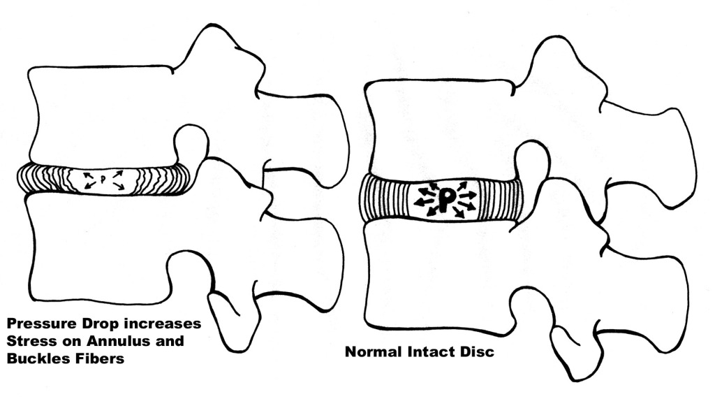
(Click to Enlarge Image) Pressure drop of nucleus causes increased stress on disc wall. The wall of the disc was designed to hold the pressurized nucleus in and to resist rotation but is not designed well to resist compression.
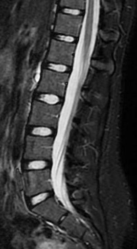
(Click to Enlarge Image) Normal MRI Discs are all well hydrated noted by the bright white color and excellent height.
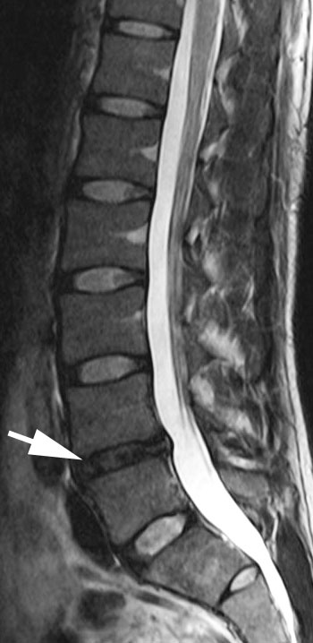
(Click to Enlarge Image) Mild Degenerative Disc Disease The arrow points to the L4-5 disc which has lost its white signal. The disc still has good height remaining and the endplates are smooth and intact.

(Click to Enlarge Image) Isolated Disc Resorption The white arrow points to the fully degenerated disc. Notice that the vertebral bodies surrouning this disc space are whiter than the other vertebra due to the leakage of fluid into the bone from the trabecular fractures.
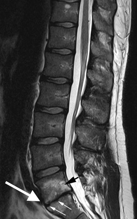
(Click to Enlarge Image) IDR The large white arrow points to the problem disc, the small white arrows to the endplate fractures and the black arrow points to the high white signal the fluid swelling inside the vertebral body.